Transitions between sleep and wakefulness are fundamental state shifts in organisms. Sleep is accompanied not only by changes in brain activity but also by widespread alterations in perception, behavior, and physiology. For example, the transition from wakefulness to sleep is associated with reductions in blood pressure, heart rate, and respiratory rate. Reciprocally, changes in peripheral physiological states can profoundly influence sleep-wake patterns and are crucial for homeostatic recovery. During infection, for instance, organisms often exhibit increased sleepiness, which helps promote recovery. How, then, are brain states and peripheral physiology coordinated?
Recently, the research teams led by Yang Dan and Yuanyuan Yao at the Institute of Neuromodulation and Cognition (INC), Shenzhen Medical Academy of Research and Translation (SMART), published a review article entitled Body-Brain Integration: The Lower Brainstem in Sleep-Wake Regulation in Annual Review of Neuroscience. The article systematically summarizes recent advances in understanding the role of the lower brainstem in sleep-wake control and proposes that sleep is not merely a brain state regulated by the central neural circuits, but also an active output of the body in response to peripheral homeostatic demands. By integrating interoceptive signals, the brainstem orchestrates a global coordination of brain states and bodily homeostasis.
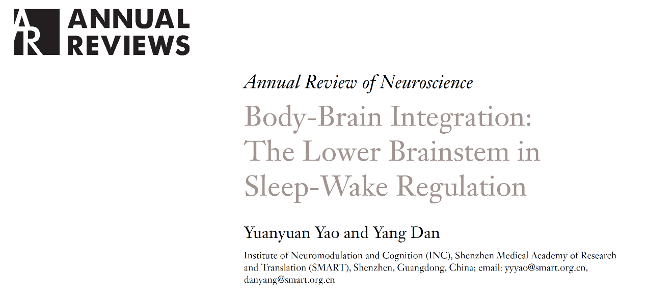
For decades, sleep research has emphasized the forebrain and hypothalamic centers as the primary regulators of sleep. However, recent studies have renewed interest in the lower brainstem. Long recognized for its roles in autonomic and somatic motor functions, the lower brainstem is now understood to be rather than a passive relay station but as an integrative hub linking internal bodily states to brain state regulation (Figure 1). It translates interoceptive signals from the cardiovascular, respiratory, digestive, and immune systems into sleep or wake drives, thereby aligning brain states with the body's homeostatic demands.
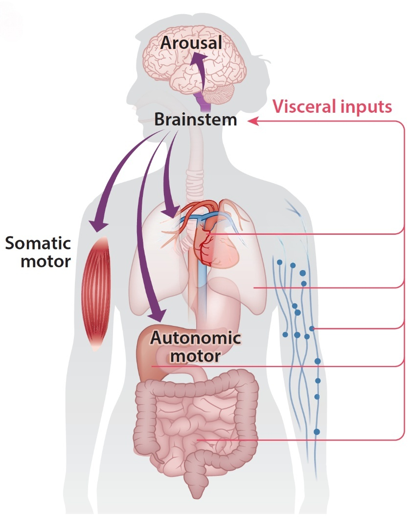
Figure 1. The brainstem is a key hub linking visceral physiology with somatic, autonomic, and brain‑state regulation.
The NST receives interoceptive signals from the cardiovascular, respiratory, and digestive systems, and detects immune‑related signals. Traditionally viewed as a homeostatic regulator of individual systems (e.g., baroreflex control of blood pressure), emerging evidence positions the NST as a crucial sleep‑regulatory hub. It translates peripheral signals such as blood pressure fluctuations and immune activation into changes in sleep drive (Figure 2), helping to align brain states with body's internal conditions. In other words, the NST is not only a major gateway for visceral sensory information entering the brain but also a key node that translates "bodily states" into "sleep need."
Figure 2. Schematic of the baroreflex circuit, with output regulating both the cardiovascular system and brain arousal.
The PBN plays a pivotal role in processing threat‑related inputs and promoting arousal. It serves as a key danger‑detection hub, integrating interoceptive inputs with somatosensory inputs such as pain and chemosensory inputs such as hypoxia to coordinate survival behaviors, and it plays a critical role in promoting arousal.
Notably, a subset of PBN neurons may also promote sleep, contributing to sickness‑induced sleep and skin-warming‑induced sleep. Future studies should explore whether other PBN functions, such as fluid intake regulation, also interact with sleep-wake control.
Sleep is not merely a change in “conscious state”; but is instead accompanied by a systemic reorganization encompassing reduced muscle tone, suppressed behavioral activity, and altered autonomic activity. GABAergic neurons in several medullary regions play a key role in coordinating these changes.
Ventromedial medulla (VMM) GABAergic neurons receive glutamatergic inputs from the sublaterodorsal nucleus in the pons, become activated during REM sleep, and mediate muscle atonia during REM sleep. They also promote NREM sleep by inhibiting monoaminergic neurons in the locus coeruleus, dorsal raphe, and ventral tegmental area, and regulate autonomic motor output via descending spinal projections.
Caudal ventrolateral medulla (CVLM) GABAergic neurons are the main inhibitory node of the arterial baroreflex. They inhibit catecholaminergic neurons in the rostral ventrolateral medulla (RVLM), reducing sympathetic outflow and vasomotor tone, and increase NREM sleep.
Parafacial zone (PZ) GABAergic neurons promote NREM sleep by inhibiting the PBN.
Although multiple subregions of the ventral medulla suppress motor activity, they have distinct effects on sleep-wake states: some promote NREM sleep, while others promote REM sleep. Systematically mapping the functional architecture of these regions and identifying the molecular and connectivity features that determine whether a given neuronal population promotes REM or NREM sleep will be essential.
Sleep is accompanied by changes in autonomic activities such as respiration and heart rate. Recent studies show that autonomic-related neuronal populations—cholinergic neurons in the nucleus ambiguus (AMB) and catecholaminergic neurons in the ventrolateral medulla and locus coeruleus (LC)—also participate in sleep-wake regulation.
Nucleus ambiguus (AMB) cholinergic neurons regulate cardiovascular, respiratory, and upper digestive functions via their projections to the heart, lungs, esophagus, pharynx, and larynx. Surprisingly, activating AMB cholinergic neurons not only lowers arterial pressure and heart rate but also promotes NREM sleep, suggesting they may help coordinate cardiovascular activity with brain state transitions.
Ventrolateral medulla (VLM) catecholaminergic neurons are activated by physiological stressors and orchestrate autonomic, metabolic, and neuroendocrine responses to promote survival. Optogenetic activation of RVLM catecholaminergic neurons rapidly induces wakefulness. Since hypoxia and hypotension - both potent activators of these neurons - signal life‑threatening conditions, the wake‑promoting effect likely facilitates immediate behavioral responses critical for survival.
The LC norepinephrine (NE) neurons integrate environmental, visceral, and cognitive inputs, and project widely throughout the brain and spinal cord, regulating behavioral states, somatic and autonomic motor activity. These neurons connect brain arousal with autonomic and somatic motor control, thereby ensuring that state transitions are matched to both external challenges and internal physiological needs.

Figure 3. Nuclei in lower brainstem involved in coordinating visceral physiology with sleep-wake regulation.
Overall, sleep is not merely a change in brain activity, but rather the result of coordinated regulation between body physiology and brain states. The lower brainstem plays an essential role in this process (Figure 3): the NST serves as the primary gateway translating visceral sensory information into sleep drive; the PBN is critical for processing threat‑related inputs and promoting arousal; catecholaminergic neurons in the VLM and LC are involved in stress‑related arousal regulation. Together with cholinergic neurons in the AMB and GABAergic neurons in multiple medullary regions,these nuclei coordinately regulate sleep-wake states, somatic motor activity, and autonomic functions. These nuclei collectively form key circuits that connect internal bodily states with brain state regulation. This framework not only provides a new theoretical basis for understanding the physiological functions of sleep, but also opens new directions for studying sleep disorders and their relationships with metabolic, immune, and cardiovascular diseases.
As technologies continue to advance and our understanding deepens, future studies are expected to systematically elucidate how distinct peripheral inputs shape the activity of specific brainstem neuronal populations to regulate sleep-wake states. These efforts will provide a more comprehensive understanding of the division of labor and coordination among brainstem nuclei in sleep regulation, while revealing the fundamental significance of body-brain integration in sleep physiology and sleep-related disorders.
Title: Body-Brain Integration: The Lower Brainstem in Sleep-Wake Regulation
Authors: Yuanyuan Yao, Yang Dan
Institute: Institute of Neuromodulation and Cognition (INC), Shenzhen Medical Academy of Research and Translation (SMART)
Journal: Annual Review of Neuroscience
Published online: April 20, 2026 (Review in Advance)
The laboratory of Yang Dan (snc@smart.org.cn) is recruiting postdoctoral fellows. The laboratory of Yuanyuan Yao (yyyao@smart.org.cn) is recruiting postdoctoral fellows, research assistants, and visiting students for graduation projects. Highly motivated applicants with backgrounds in neuroscience or related disciplines are warmly welcome to join us.
Translation: Jianming JIA
Proofreading: Fanfan SUN
talent@smart.org.cn
researcher@smart.org.cn
recruitment@smart.org.cn
graduate_office@smart.org.cn
graduate_admission@smart.org.cn
otl@smart.org.cn
smartfund@smart.org.cn
pr@smart.org.cn
Subscription successful! Thank you for following SMART.