Glycans are one of the four fundamental biological macromolecules and play essential roles in a wide range of physiological processes, including cell recognition, signal transduction, immune regulation, and structural support. Historically, glycans were often collectively referred to as "carbohydrates," a term derived from their classical chemical formula, Cx(H2O)n. Although glycan research can be traced back to the 19th century, the field has long lagged behind protein and nucleic acid research. A fundamental reason is the extreme complexity of glycans and the longstanding lack of effective analytical approaches.
Unlike DNA, RNA, and proteins, glycan biosynthesis is not template-driven and therefore lacks a direct genome-to-sequence correspondence. In addition, the enormous diversity of monosaccharide composition, complex glycosidic linkages, and substantial conformational heterogeneity have greatly limited high-resolution three-dimensional structural analysis of complex glycan assemblies. The long-standing absence of structural information has also hindered a deeper understanding of the roles of glycans in diverse physiological and pathological processes.
Figure 1. The online homepage of the article.
On April 23, 2026, Nieng Yan, Junhao Huang, Chuangye Yan, and colleagues published a research article in Science entitled "Structural N- and O-glycans revealed by high-resolution cryo-EM analysis of tubular mastigonemes". Using tubular mastigonemes (T-mastigonemes) from the chrysophyte Ochromonas danica as the research subject, the researchers established an integrated strategy combining high-resolution cryo-EM, glycoproteomics, and AI modeling. The study achieved cryo-EM density maps with an overall glycan resolution of approximately 2 Å and local core resolutions reaching 1.8 Å, advancing the structural analysis of natural glycan complexes to near-atomic resolution.
This level of resolution enabled direct visualization of natural glycans, allowing accurate identification of multiple monosaccharide types as well as unambiguous assignment of covalent modifications including acetylation, methylation, and sulfation. By integrating structural biology with targeted mass spectrometry, the researchers further identified a novel non-canonical N-glycosylation type based on an "AND" motif and revealed its evolutionary conservation in multiple highly pathogenic species through structural clustering analyses.
Meanwhile, the AI-based automated modeling tool EModelG, developed by the Nieng Yan laboratory, substantially improved the efficiency of glycan structure modeling, enabling a transition from indirect fragment-based inference to high-precision direct modeling and providing methodological support for large-scale structural studies of complex natural glycans.
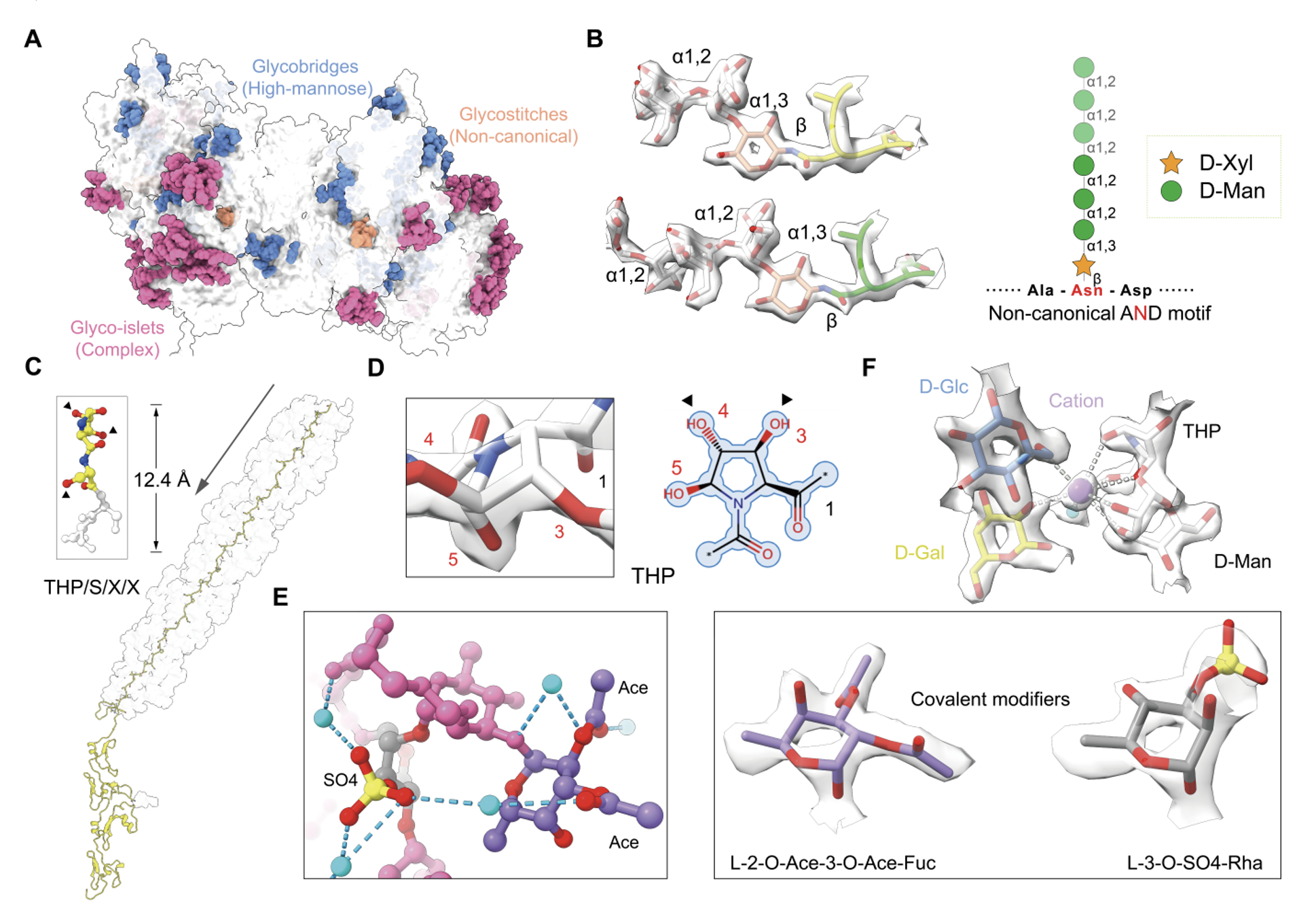
Figure 2. High-resolution cryo-EM analysis reveals structural N- and O-glycans in tubular mastigonemes
Further structural analyses showed that different classes of N-glycans perform distinct structural functions within the assembly. The study systematically proposed three glycan-mediated modes of macromolecular assembly.
First, "glycostitch" structures formed by glycans attached to non-canonical AND motifs adopt regular helical conformations that stabilize subunit interfaces within repeating structural units. Second, "glycobridges," primarily mediated by high-mannose glycans, span neighboring repeating units to establish long-range interactions and exhibit a degree of evolutionary conservation across species. Third, "glyco-islets" are distributed as clustered surface patches that enhance overall stability and solubility by increasing surface area and hydrophilicity.
These findings challenge the conventional view of glycans as merely accessory modifications on proteins and demonstrate that glycans themselves can directly function as key structural elements driving high-order biomolecules assembly.
In terms of O-glycan structures, the researchers further resolved high-order glycofibrils formed by PSXX tetrapeptide repeats. The structures revealed dense glycosylation at 3,4,5-trihydroxyproline (THP) and serine residues, generating regularly arranged surface "glycan-spike" protrusions that collectively form a stable glycofibril scaffold. The study represents the first identification of THP modification within this system and precisely resolved a complex O-glycan folding pattern composed of eight monosaccharide types together with multiple covalent modifications.
More importantly, the high-resolution density maps also revealed extensive ordered water-molecule networks and multivalent cation coordination centers. These structural features, which were previously difficult to observe directly, significantly strengthened glycan–glycan interactions through hydrogen-bonding networks and coordination effects. In particular, metal cations were found to mediate eight-coordinate networks spanning neighboring repeating units, thereby enhancing the mechanical rigidity of the glycan helical core. This process is accompanied by characteristic mannose "twisted-boat" conformational flips that provide local energetic compensation, offering direct experimental evidence for the physicochemical basis underlying the stability of complex glycan assemblies.
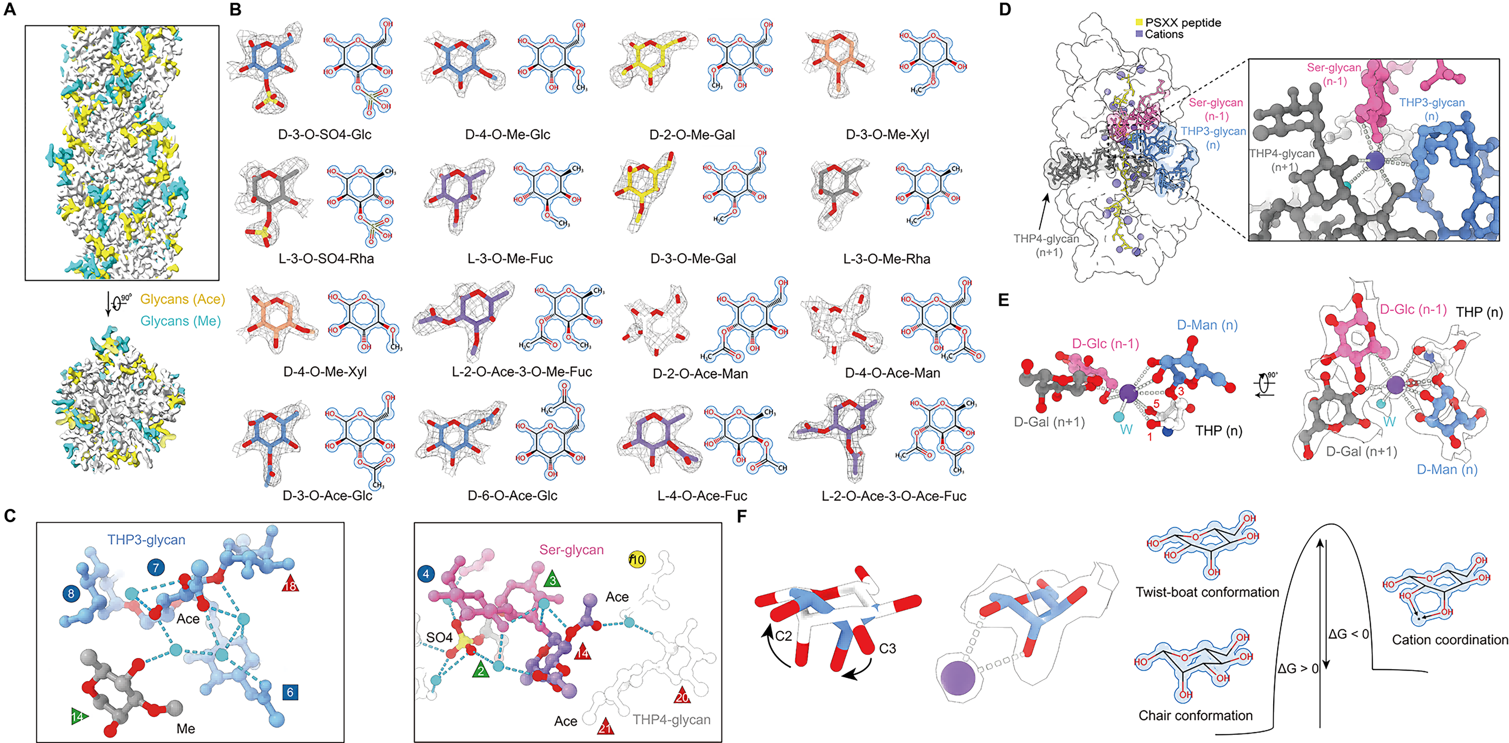
Figure 3. Structural roles of covalent modifications, water molecules, and metal ions in glycan helical assembly
Taken together, this study achieved direct structural characterization of natural glycans from chemical composition to three-dimensional conformation. The researchers proposed a series of new glycan assembly elements, including "glycostitch," "glycobridge," and "glyco-islet," and systematically revealed the structural roles of glycans in high-order biomolecular assembly. These findings provide a new methodological framework for the discovery, characterization, and engineering of complex glycan architectures, marking the entry of glycan structural biology into an era of atomic-resolution analysis.
Nieng Yan, Founding President of Shenzhen Medical Academy of Research and Translation (SMART) and Director of Shenzhen Bay Laboratory; Junhao Huang, Ph.D. at the School of Life Sciences, Tsinghua University; and Associate Professor Chuangye Yan are the co-corresponding authors of the study. Junhao Huang, ShuiMu Scholar Hui Tao, and Sheng Chen are the co-first authors. Research Assistant Yahua Cui and Ph.D. student Yiran Xu also made important contributions to this work.
Cryo-EM data collection and computational analyses were supported by the Biological Structure Analysis Platform and High-Performance Computing Platform of SMART. Mass spectrometry analyses were supported by the Proteomics and Protein Chemistry Platform of Tsinghua University and by Junior Principal Investigator Xinlei Sheng from the Institute of Human Immunology at SMART.
This study was initially released on January 28, 2026, through the LTS Preprint Server (LTSpreprints.org)[1], where it was made publicly available for reading, discussion, and scientific exchange prior to formal publication, while also serving as an important reference for subsequent studies. Articles posted on LTSpreprints are increasingly being indexed by major academic platforms such as Google Scholar and ResearchGate, reflecting their growing academic impact. The platform is dedicated to fostering an open, efficient, and standardized environment for scientific communication by providing rapid dissemination, broad visibility, and long-term archiving for research outputs.
This work was supported by the Major Research Plan "Decoding the Sugar Code of Life" of the National Natural Science Foundation of China, the NSFC Youth Student Basic Research Project, the Beijing Frontier Research Center for Biological Structure, and the Tsinghua-Peking Joint Center for Life Sciences. The team also expressed special thanks for support provided through the "Mindray Professorship" funded by the Pengrui Foundation.
The glycofibril structures and corresponding cryo-EM density maps reported in this study have been deposited in the CryoSeek Database (see references [2] and [3] for structural models and density maps). Established by the SMART under the leadership of Nieng Yan, the CryoSeek Database systematically archives structural and identification data related to biological "dark matter," particularly glycans, and provides online services to facilitate data management and sharing within the research community. Related studies describing the CryoSeek Database and the high-throughput CryoSeek structural determination platform have also been released on LTS Preprint Server[4].
Reference:
[1] Huang, J., Tao, H., Chen, S., Cui, Y., Xu, Y., Yan, C., & Yan, N. (2026). AI-facilitated high-resolution cryo-EM analyses of tubular mastigonemes reveal the structural roles of N- and O-glycans. LangTaoSha Preprint Server. https://doi.org/10.65215/LTSpreprints.2026.01.28.000106
[2]https://www.cryoseek.org.cn/home/maps/20260421173015A321/
[3]https://www.cryoseek.org.cn/home/maps/20260421172519A305/
[4] Hu, M., Chen, S., Wang, T., Qin, L., Zhang, Q., Zhang, Y., Ge, Q., Chen, T., Li, M., Li, C., Xu, G., Gui, Q., Li, Z., & Yan, N. (2025). High-throughput cryo-EM characterization and automated model building of glycofibrils via CryoSeek. LangTaoSha Preprint Server. https://doi.org/10.65215/bkvrt910
Translation: Jianming JIA
talent@smart.org.cn
researcher@smart.org.cn
recruitment@smart.org.cn
graduate_office@smart.org.cn
graduate_admission@smart.org.cn
otl@smart.org.cn
smartfund@smart.org.cn
pr@smart.org.cn
Subscription successful! Thank you for following SMART.