Carbohydrates play indispensable roles in fundamental biological processes, ranging from energy metabolism and cell signaling to immune recognition. These diverse functions are rooted in their unique three-dimensional (3D) stereochemical structures. However, compared to nucleic acids and proteins, the structural and functional study of glycans has lagged significantly. The primary obstacles include the extraordinary diversity of monosaccharides, multiple chiral centers, complex glycosidic linkages in polysaccharides, and the intrinsic conformational flexibility of glycan structures. These factors collectively make high-resolution 3D structural determination exceptionally difficult, limiting our mechanistic understanding of glycan biology.
To address these challenges, the team led by Nieng Yan proposed the CryoSeek strategy. This approach utilizes cryogenic electron microscopy (cryo-EM) as a discovery tool for previously unknown biomolecular entities, pioneering a "forward structural biology" paradigm centered on structure-first discovery[1–4]. By analyzing environmental samples from the Tsinghua Lotus Pool (TLP), the researchers successfully identified a series of "glycofibrils." These studies not only highlight the central role of glycans in biomolecular assembly but also establish a multidisciplinary framework for high-throughput structural analysis and sequencing of natural glycans.
Subsequently, the teams of Mingxu Hu and Nieng Yan collaborated to enhance the throughput of the CryoSeek platform. By developing large-scale image classification algorithms and AI-based glycan modeling tools, they achieved the structural characterization of numerous environmental glycofibrils and established the CryoSeek Database[5].
Despite these advances, cryo-EM structural determination faces a fundamental hurdle: the loss of "absolute hand" (handedness) information during 2D image acquisition. Consequently, reconstructed 3D density maps often exist in two mirror-related (enantiomeric) forms. For proteins, this ambiguity is easily resolved because α-helices are almost universally right-handed. However, glycofibrils—composed primarily of carbohydrates—lack such internal biological markers. Furthermore, since both D- and L-type sugars occur in nature, determining the absolute hand of a glycofibril remains a major challenge even at atomic resolution. This uncertainty is akin to losing the "North-South" orientation on a map, making accurate atomic modeling nearly impossible.
Traditionally, this has been addressed via "tilt-pair" imaging[6], which requires prior structural knowledge and repeated imaging of the same field at two different tilt angles. This process is technically demanding, time-consuming, and expensive. More importantly, it is impractical for highly heterogeneous environmental samples, which may contain hundreds of distinct species in a single natural water sample.
On February 26, 2026, Mingxu Hu, Jiawei Wang, Nieng Yan, and their collaborators published a research article titled "Absolute hand determination of glycofibrils from natural sources in cryo-EM" in the Proceedings of the National Academy of Sciences (PNAS).
To overcome this bottleneck, the researchers developed Ahaha (Absolute hand determination of helical assembly). Using only conventional cryo-EM imaging under a single tilted condition, Ahaha efficiently and accurately determines the absolute hand of naturally derived glycofibrils. The method is now accessible via an online service (Figure 1).

Figure 1. Interface of the Ahaha Online Service (https://cryoseek.org/ahaha)
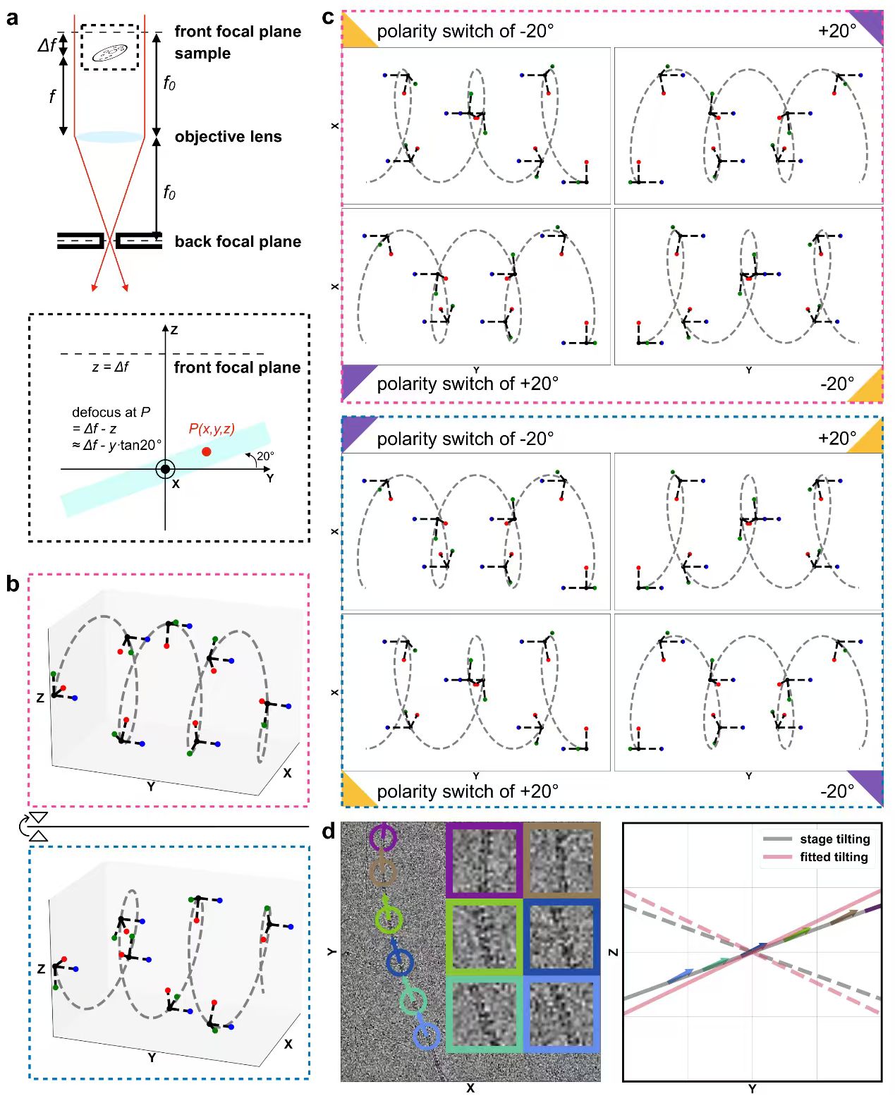
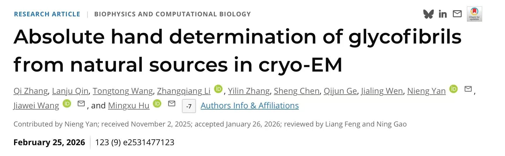
Figure 2. Schematic diagram illustrating the principle behind Ahaha's determination of chirality.
The principle of Ahaha is illustrated in Figure 2. enantiomeric helical structures (Figure 2b) generate different projections under the same tilt angle (for example, +20°), but produce identical projections under opposite tilt angles (for example, +20° versus −20°) (Figure 2c). Therefore, when the same projection image is observed, helices with opposite handedness must correspond to opposite tilt directions.
On the other hand, because helical glycofibrils naturally lie flat on the specimen plane, their tilt direction must be aligned with the specimen tilt itself. By analyzing whether the particle tilt angles obtained during high-resolution helical reconstruction are consistent with the experimentally applied specimen tilt direction, Ahaha can determine whether the handedness of the reconstructed high-resolution density map is correct (Figure 2d).
When the particle tilt angles are approximately parallel to the assigned specimen tilt direction, the handedness of the reconstructed helix is considered correct. Conversely, if the particle tilt angles are oriented oppositely to the specimen tilt direction, the reconstructed helix is regarded as having the opposite hand.
In addition, as a complementary validation step in Ahaha analysis, particle defocus parameters (e.g., from CryoSPARC Patch CTF) can be used to cross-validate whether the assigned specimen tilt direction is correct (Figure 2a).
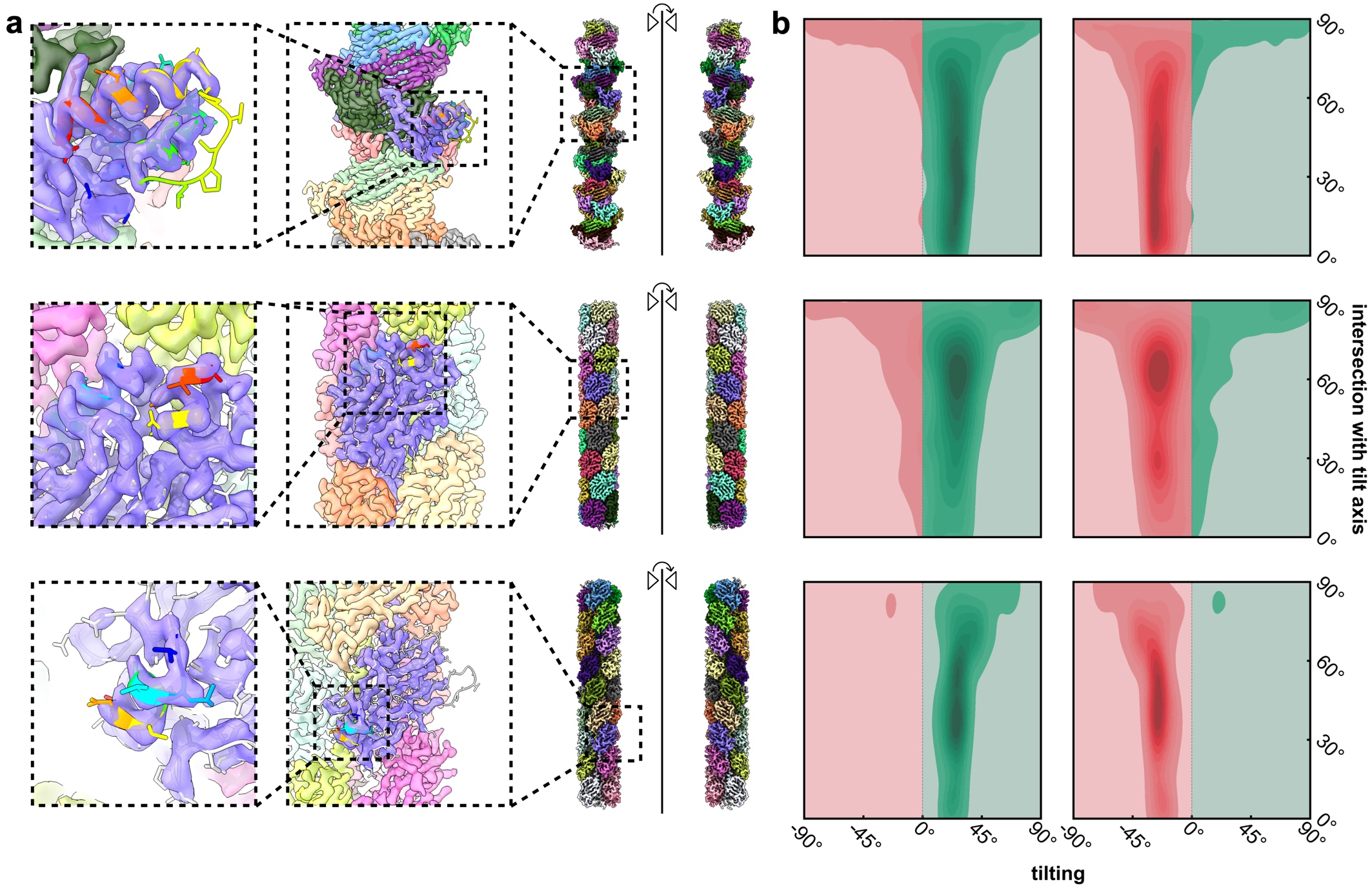
Figure 3. The validity of Ahaha was verified using pilus-like helical structures.
To validate the accuracy of Ahaha, the researchers analyzed three pilus-like structures identified in a natural freshwater sample collected from stalactite drip water in a karst cave. The three rows in Figure 3 correspond to the helical structures termed pilus-like-α, pilus-like-β, and pilus-like-γ, respectively. These assemblies are primarily composed of proteins.
The reconstructed density maps and their enantiomeric counterparts are shown in Figure 3a. Among them, the structures on the left possess the correct absolute hand, as the α-helices in proteins are known to be right-handed. Enlarged views of the α-helical regions within the density maps are shown together with the fitted atomic models.
The Ahaha analysis results are presented in Figure 3b. Ahaha generates heat maps based on the particle tilt angles and the angular relationship between particle orientations and the tilt axis, while simultaneously evaluating whether the particle tilt directions are consistent with the assigned specimen tilt direction. The heat maps on the left and right correspond to density maps with correct and incorrect handedness, respectively.
Positive tilt values (green) are aligned with the specimen tilt direction, whereas negative tilt values (red) are oriented opposite to the specimen tilt direction. For reconstructions with the correct absolute hand, particles in the Ahaha heat maps are predominantly distributed within the green regions, indicating that Ahaha correctly determines the handedness of the helical structures. In contrast, for reconstructions with the opposite hand, particle distributions are concentrated in the red regions, indicating that Ahaha correspondingly assigns the opposite handedness.
Figure 4. (A) Use Ahaha to determine the absolute chirality of four naturally occurring carbohydrate fibers. For a pair of enantiomeric helical structures, the dominance of green regions in the heatmap provided by Ahaha indicates that Ahaha correctly determined the absolute chirality of the structure. (B) Structures with correct absolute chirality, along with their adjacent helical units and helical symmetry parameters.
Using Ahaha, the researchers successfully determined the absolute handedness of four glycofibrils identified from the same natural freshwater sample (Figure 4A). As a control, enantiomeric structures with opposite absolute handedness were intentionally generated during helical reconstruction by using enantiomeric initial models together with opposite helical twist parameters. Correspondingly, Ahaha produced opposite handedness assignments for these reconstructions, further validating the accuracy of the method.
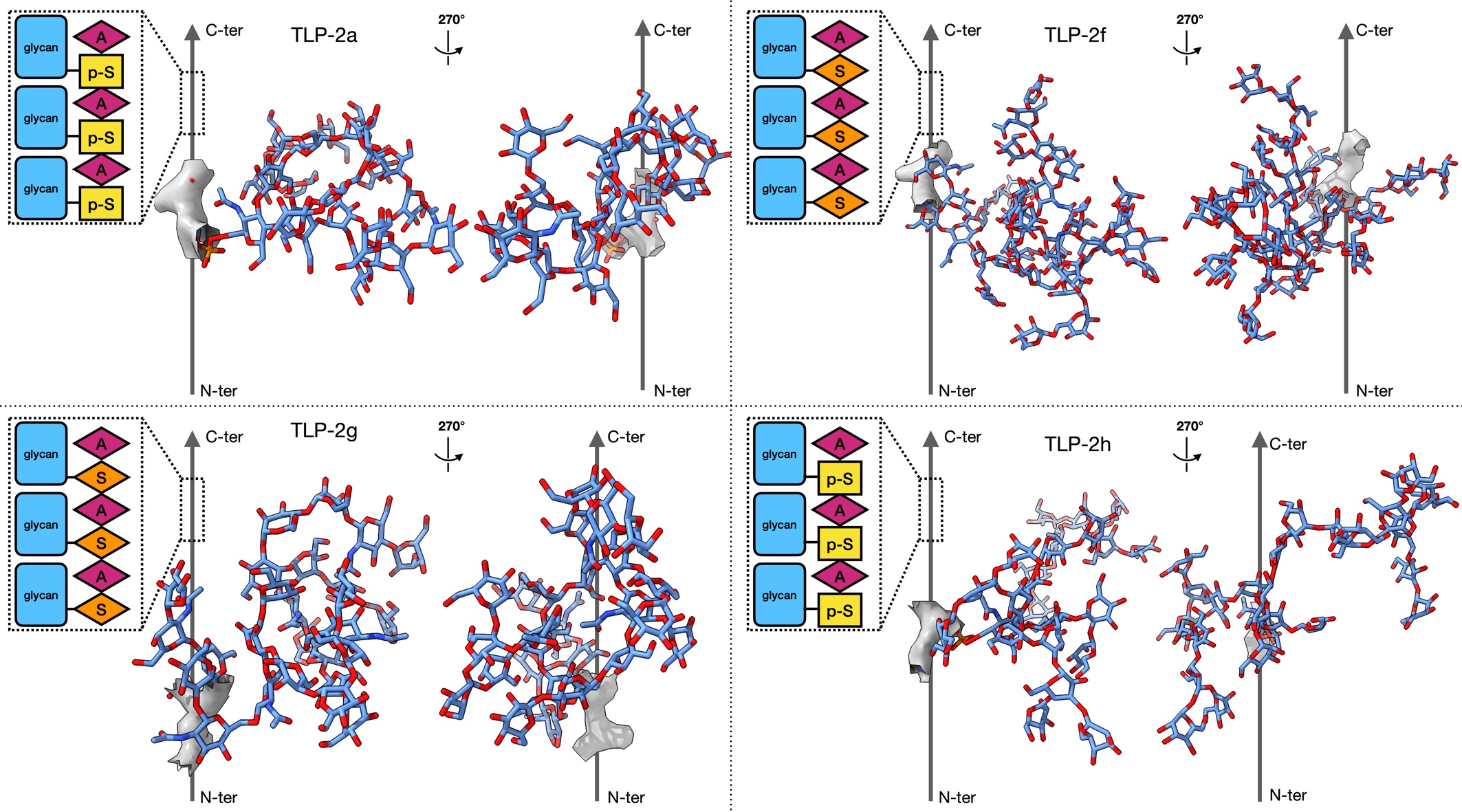
Figure 5. Atomic models of the glycofibrils.
Based on the Ahaha analysis, the researchers further built atomic models for the glycofibrils (Figure 5). The models reveal monosaccharide chains linked to serine or phosphoserine residues. Gray arrows indicate linear peptide chains composed of repeating dipeptide motifs. The N- and C-termini were inferred from the positions of oxygen atoms in amino acid carboxyl groups, although definitive assignment will require higher-resolution density maps.
The schematic diagrams within the dashed boxes illustrate the composition of the linear peptide chains and the corresponding O-glycosylation sites. "SA" is used as a general notation representing possible dipeptide combinations of serine/threonine and alanine/glycine residues. Due to the current resolution limit, the glycan identities included in the models represent the best approximations under the available experimental conditions. When the density maps did not provide sufficient evidence for assigning specific sugar types, mannose was used as the default glycan model.
These structural models mark an important step toward elucidating the architecture of glycans, often referred to as the "dark matter" of life, and may help accelerate future breakthroughs in glycobiology.
Mingxu Hu, Junior PI at Shenzhen Medical Academy of Research and Translation (SMART) ; Jiawei Wang, Associate Professor at the School of Life Sciences, Tsinghua University; and Nieng Yan, Founding Dean of Shenzhen Medical Academy of Research and Translation and Director of Shenzhen Bay Laboratory, are the co-corresponding authors of the study. Dr. Qi Zhang from SMART is the first author. Qin Lanju, Dr. Tongtong Wang, Assistant Researcher Zhangqiang Li, Yilin Zhang, Dr. Sheng Chen, Qijun Ge, and Jialing Wen also made important contributions to this work.
This research was supported by Shenzhen Medical Academy of Research and Translation, the National Natural Science Foundation of China, and the Beijing Frontier Research Center for Biological Structure.
Reference:
[1] Wang, T., Li, Z., Xu, K., Huang, W., Huang, G., Zhang, Q. C., & Yan, N. (2024). CryoSeek: A strategy for bioentity discovery using cryoelectron microscopy. Proceedings of the National Academy of Sciences, 121(42), e2417046121.
[2] Wang, T., Huang, W., Xu, K., Sun, Y., Zhang, Q.C., Yan, C., Li, Z., & Yan, N. (2025). CryoSeek II: Cryo-EM analysis of glycofibrils from freshwater reveals well-structured glycans coating linear tetrapeptide repeats, Proceedings of the National Academy of Sciences, 122(1), e2423943122.
[3] Wang, T., Sun, Y., Li, Z., & Yan, N. (2024). The 8-nm spaghetti: well-structured glycans coating linear tetrapeptide repeats discovered from freshwater with CryoSeek. bioRxiv, 2024-12.
[4] Li, Z., Wang, T., Sun, Y., Xu, K., Huang, W., Zhang, Q., Yan. C., Hu, M., Yan, N. (2024) CryoSeek identification of glycofibrils with diverse compositions and structural assemblies. bioRxiv, 2025-10.
[5] Hu, M., Chen, S., Wang, T. , Qin, L. , Zhang, Q. , Zhang, Y., Ge, Q., Chen, T., Li, M., Li, C., Xu, G., Gui, Q., Li, Z. , Yan, N. (2025). CryoSeek identification of glycofibrils with diverse compositions and structural assemblies. LTS Preprint Server, 2025-11.
[6] Rosenthal, P., & Henderson, R. (2003). Optimal Determination of Particle Orientation, Absolute Hand, and Contrast Loss in Single-particle Electron Cryomicroscopy. Journal of Molecular Biology, 333(4), 721–745, 2023-10.
Translation: Jianming JIA
talent@smart.org.cn
researcher@smart.org.cn
recruitment@smart.org.cn
graduate_office@smart.org.cn
graduate_admission@smart.org.cn
otl@smart.org.cn
smartfund@smart.org.cn
pr@smart.org.cn
Subscription successful! Thank you for following SMART.